the eye and its primary function is to control the
amount of light hitting the retina.
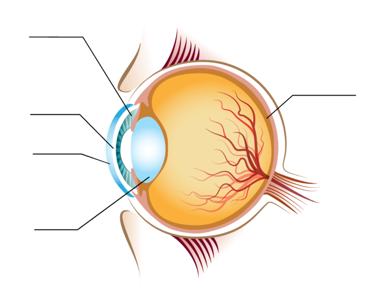
the Iris. The iris adjusts the size
of the pupil and controls the amount
of light that can enter inside.
The cornea is the clear front surface of the eye.
On cross section, the cornea contains five distinct layers;
- Epithelium
- Bowman’s Layer
- Stroma
- Descemet’s membrance
- Endothelium
The outside surface layer is composed of epithelial cells that are easily abraded.
Though epithelial injuries are painful, this layer heals quickly and typically does not scar.
Under this lies Bowman’s layer and then the stroma. The corneal stroma makes up 90% of the corneal
thickness, and if the stroma is damaged this can lead to scar formation.
The next layer is Descemet’s membrane, which is really the basal lamina of the endothelium, the final inner layer.
The inner endothelium is only one cell layer thick and works as a pump to keep the cornea dehydrated.
It gets its nourishment entirely from nutrients
floating in the aqueous fluid. The lens
also has the highest protein concentration of
any tissue in the body (65% water,
35% protein).Human lens contains Lutein and
Zeaxanthin for protection from sunlight.
Due to age, proteins begin to
break down and the lens get cloudy
leading to clouded vision. This condition
is known as Cataract.
The retina is the sensory portion of the eye and contains layers of photoreceptors, nerves, and supporting cells.
Light must travel through the thickness of the retina before striking and activating the rods & cones. Subsequently the absorption of photons by the visual pigment of the photoreceptors is translated into first a biochemical message and then an electrical message that can stimulate all the succeeding neurons of the retina.
Central Retina & Peripheral Retina
Central retina close to the fovea is considerably thicker than peripheral retina This is due to the increased packing density of photoreceptors, particularly the cones,
Central retina is cone-dominated retina whereas peripheral retina is rod-dominated.
Macula / Macula Lutea & Foveal pit.
The macula or macula lutea (from Latin macula, "spot" + lutea, "yellow") is an oval-shaped highly pigmented yellow spot near the center of the retina of the human eye. It has a diameter of around 5 mm. Near its
center is the fovea, a small pit that contains the largest concentration of cone cells in the eye and is
responsible for central, high resolution vision.